RET (タンパク質)
RETはGDNFファミリーの細胞外シグナル伝達分子を結合する受容体型チロシンキナーゼであり、ヒトではRET遺伝子にコードされる[5]。RET遺伝子の機能喪失型変異はヒルシュスプルング病の発症と関係しており[6]、機能獲得型変異は甲状腺髄様癌、多発性内分泌腺腫症2A型と2B型を含む、さまざまなタイプのがんの発症と関係している[7]。
構造
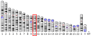
[編集]RETは"rearranged during transfection"の略であり、この遺伝子のDNA配列はもともと、3T3線維芽細胞株をヒトリンパ腫細胞由来のDNAでトランスフェクションした際に再編成が起こっている領域として発見された[8]。ヒトのRET遺伝子は10番染色体(10q11.2)に位置しており、21個のエクソンからなる[9]。
RET遺伝子からは選択的スプライシングによって3つの異なるタンパク質アイソフォームが産生される。RET51、RET43、RET9はC末端のテールがそれぞれ51、43、9アミノ酸からなる[10]。一般的なアイソフォームはRET51とRET9で、in vivoでの生物学的役割が最もよく研究されている。
各アイソフォームは共通のドメイン構造を持ち、4つのカドヘリン様リピートとシステインリッチ領域からなるN末端の細胞外ドメイン、疎水的な膜貫通ドメイン、細胞質側のチロシンキナーゼドメインから構成される。チロシンキナーゼドメインは27アミノ酸からなる挿入配列によって分割されている。RET9、18、51のチロシンキナーゼドメイン内には16個のチロシン(Tyr)が存在する。Tyr1090とTyr1096はRET51アイソフォームにのみ存在する[11]。
RETの細胞外ドメインには9つのN-グリコシル化部位が存在する。完全にグリコシル化されたRETタンパク質は170 kDaであるとの報告があるが、どのアイソフォームに対応するものであるかは明らかではない[12]。
キナーゼの活性化
[編集]RETはGDNFファミリーリガンド(GFL)に対する受容体である[13]。
RETを活性化するためには、GFLはまずGPIアンカーで膜に固定されたコレセプターと複合体を形成する必要がある。コレセプター自身はGFRα(GDNF receptor-α)タンパク質ファミリーに分類される。さまざまなGFRαファミリーのメンバー(GFRα1、GFRα2、GFRα3、GFRα4)は、それぞれ特定のGFLに対して特異的な結合活性を示す[14]。GFL-GFRα複合体が形成されると、複合体は2つのRET分子を結合させ、各RET分子のチロシンキナーゼドメイン内の特定のチロシン残基のトランス自己リン酸化を開始させる。キナーゼドメインの活性化ループ(Aループ)に位置するTyr900とTyr905が自己リン酸化部位であることは質量分析によって示されている[15]。Tyr905のリン酸化はキナーゼの活性型コンフォメーションを安定化し、主にC末端のテール領域に位置する他のチロシン残基の自己リン酸化を引き起こす[11]。
発生におけるRETシグナル伝達の役割
[編集]GDNF、GFRα1またはRETタンパク質自身を欠損したマウスは、腎臓と腸管神経系の発生に重大な欠陥が生じる。このことは、RETシグナルの伝達が腎臓と腸管神経系の正常な発生に重要であることを示唆している[11]。
臨床的意義
[編集]RETの活性化型点変異によって、多発性内分泌腫瘍症2型(MEN2)と呼ばれる遺伝性がん症候群が生じる[16]。臨床症状によって、MEN2A、MEN2B、家族性甲状腺髄様癌(FMTC)の3つのサブタイプが存在する[7]。点変異の位置と疾患の表現型の間には高度の相関がみられる。
染色体再編成によってRETタンパク質のC末端領域が他のタンパク質のN末端部分に並置された融合遺伝子が生じることで、RETのキナーゼ活性の恒常的な活性化が引き起こされることがある。このようなタイプの変異は甲状腺乳頭癌と関係しており、形成される融合がんタンパク質はRET/PTCタンパク質と呼ばれている[17]。
疾患データベース
[編集]ユタ大学のRET遺伝子の変異データベースでは、2020年1月時点で199種類の変異が同定されている。
相互作用
[編集]RETは次に挙げる因子と相互作用することが示されている。
出典
[編集]- ^ a b c GRCh38: Ensembl release 89: ENSG00000165731 - Ensembl, May 2017
- ^ a b c GRCm38: Ensembl release 89: ENSMUSG00000030110 - Ensembl, May 2017
- ^ Human PubMed Reference:
- ^ Mouse PubMed Reference:
- ^ “Structure and chemical inhibition of the RET tyrosine kinase domain”. J. Biol. Chem. 281 (44): 33577–87. (2006). doi:10.1074/jbc.M605604200. PMID 16928683.
- ^ Martucciello, G.; Ceccherini, I.; Lerone, M.; Jasonni, V. (2000-07). “Pathogenesis of Hirschsprung's disease”. Journal of Pediatric Surgery 35 (7): 1017–1025. doi:10.1053/jpsu.2000.7763. ISSN 0022-3468. PMID 10917288.
- ^ a b “RET germline mutations identified by exome sequencing in a Chinese multiple endocrine neoplasia type 2A/familial medullary thyroid carcinoma family”. PLoS ONE 6 (5): e20353. (2011). Bibcode: 2011PLoSO...620353Q. doi:10.1371/journal.pone.0020353. PMC 3105051. PMID 21655256.
- ^ “Activation of a novel human transforming gene, ret, by DNA rearrangement.”. Cell 42 (2): 581–8. (1985). doi:10.1016/0092-8674(85)90115-1. PMID 2992805.
- ^ “Exon structure and flanking intronic sequences of the human RET proto-oncogene”. Biochem. Biophys. Res. Commun. 196 (3): 1288–1295. (1993). doi:10.1006/bbrc.1993.2392. PMID 7902707.
- ^ “Characterization of RET proto-oncogene 3' splicing variants and polyadenylation sites: a novel C-terminus for RET”. Oncogene 11 (10): 2039–2045. (1995). PMID 7478523.
- ^ a b c “RET tyrosine kinase signaling in development and cancer”. Cytokine Growth Factor Rev. 16 (4–5): 441–67. (2005). doi:10.1016/j.cytogfr.2005.05.010. PMID 15982921.
- ^ “Characterization of the ret proto-oncogene products expressed in mouse L cells”. Oncogene 8 (11): 2925–2929. (1993). PMID 8414495.
- ^ “The GDNF family ligands and receptors - implications for neural development”. Curr. Opin. Neurobiol. 10 (1): 103–10. (2000). doi:10.1016/S0959-4388(99)00048-3. PMID 10679429.
- ^ “GDNF family neurotrophic factor signaling: four masters, one servant?”. Mol. Cell. Neurosci. 13 (5): 313–25. (1999). doi:10.1006/mcne.1999.0754. PMID 10356294.
- ^ “Identification of RET autophosphorylation sites by mass spectrometry”. J. Biol. Chem. 279 (14): 14213–24. (2004). doi:10.1074/jbc.M312600200. PMID 14711813.
- ^ Online 'Mendelian Inheritance in Man' (OMIM) MULTIPLE ENDOCRINE NEOPLASIA, TYPE IIA; MEN2A -171400
- ^ Menicali, Elisa; Moretti, Sonia; Voce, Pasquale; Romagnoli, Serena; Avenia, Nicola; Puxeddu, Efisio (2012). “Intracellular signal transduction and modification of the tumor microenvironment induced by RET/PTCs in papillary thyroid carcinoma”. Frontiers in Endocrinology 3: 67. doi:10.3389/fendo.2012.00067. ISSN 1664-2392. PMC 3357465. PMID 22661970.
- ^ “Role of Dok1 in cell signaling mediated by RET tyrosine kinase”. J. Biol. Chem. 277 (36): 32781–90. (September 2002). doi:10.1074/jbc.M202336200. PMID 12087092.
- ^ “Dok-6, a Novel p62 Dok family member, promotes Ret-mediated neurite outgrowth”. J. Biol. Chem. 279 (40): 42072–81. (October 2004). doi:10.1074/jbc.M403726200. PMID 15286081.
- ^ “Novel p62dok family members, dok-4 and dok-5, are substrates of the c-Ret receptor tyrosine kinase and mediate neuronal differentiation”. J. Cell Biol. 154 (2): 345–54. (July 2001). doi:10.1083/jcb.200102032. PMC 2150770. PMID 11470823.
- ^ “A GPI-linked protein that interacts with Ret to form a candidate neurturin receptor”. Nature 387 (6634): 717–21. (June 1997). doi:10.1038/42722. PMID 9192898.
- ^ “Binding of GDNF and neurturin to human GDNF family receptor alpha 1 and 2. Influence of cRET and cooperative interactions”. J. Biol. Chem. 275 (36): 27505–12. (September 2000). doi:10.1074/jbc.M000306200. PMID 10829012.
- ^ a b “The Ret receptor protein tyrosine kinase associates with the SH2-containing adapter protein Grb10”. J. Biol. Chem. 270 (37): 21461–3. (September 1995). doi:10.1074/jbc.270.37.21461. PMID 7665556.
- ^ “Direct association between the Ret receptor tyrosine kinase and the Src homology 2-containing adapter protein Grb7”. J. Biol. Chem. 271 (18): 10607–10. (May 1996). doi:10.1074/jbc.271.18.10607. PMID 8631863.
- ^ a b “The oncogenic versions of the Ret and Trk tyrosine kinases bind Shc and Grb2 adaptor proteins”. Oncogene 9 (6): 1661–8. (June 1994). PMID 8183561.
- ^ “Identification of Shc docking site on Ret tyrosine kinase”. Oncogene 14 (7): 773–82. (February 1997). doi:10.1038/sj.onc.1200896. PMID 9047384.
- ^ “Central role of the threonine residue within the p+1 loop of receptor tyrosine kinase in STAT3 constitutive phosphorylation in metastatic cancer cells”. Mol. Cell. Biol. 24 (21): 9390–400. (November 2004). doi:10.1128/MCB.24.21.9390-9400.2004. PMC 522220. PMID 15485908.
- ^ “Activation of signal transducer and activator of transcription 3 by oncogenic RET/PTC (rearranged in transformation/papillary thyroid carcinoma) tyrosine kinase: roles in specific gene regulation and cellular transformation”. Mol. Endocrinol. 17 (6): 1155–66. (June 2003). doi:10.1210/me.2002-0401. PMID 12637586.
- ^ “MEN2A-RET-induced cellular transformation by activation of STAT3”. Oncogene 20 (38): 5350–8. (August 2001). doi:10.1038/sj.onc.1204715. PMID 11536047.
関連文献
[編集]- “Mutations of the RET proto-oncogene in the multiple endocrine neoplasia type 2 syndromes, related sporadic tumours, and hirschsprung disease”. Hum. Mutat. 9 (2): 97–109. (1997). doi:10.1002/(SICI)1098-1004(1997)9:2<97::AID-HUMU1>3.0.CO;2-M. PMID 9067749.
- “Mutations in Hirschsprung disease: when does a mutation contribute to the phenotype”. Eur. J. Hum. Genet. 5 (4): 180–5. (1998). PMID 9359036.
- “RET/PTC rearrangement in thyroid tumors”. Endocr. Pathol. 13 (1): 3–16. (2002). doi:10.1385/EP:13:1:03. PMID 12114746.
- “Minireview: RET: normal and abnormal functions”. Endocrinology 145 (12): 5448–5451. (2004). doi:10.1210/en.2004-0922. PMID 15331579.
- “Dysfunction of the RET receptor in human cancer”. Cell. Mol. Life Sci. 61 (23): 2954–2964. (2005). doi:10.1007/s00018-004-4276-8. PMID 15583857.
- “[RET mutations and preventive treatment of medullary thyroid cancer]”. Ann. Endocrinol. 66 (3): 168–75. (2005). doi:10.1016/s0003-4266(05)81748-2. PMID 15988377.
- “Molecular mechanisms of RET-induced Hirschsprung pathogenesis”. Ann. Med. 38 (1): 11–9. (2006). doi:10.1080/07853890500442758. PMID 16448984.
- “RET/PTC rearrangements and BRAF mutations in thyroid tumorigenesis”. Endocrinology 148 (3): 936–41. (2007). doi:10.1210/en.2006-0921. PMID 16946010.
- “Current concepts in RET-related genetics, signaling and therapeutics”. Trends Genet. 22 (11): 627–36. (2007). doi:10.1016/j.tig.2006.09.005. PMID 16979782.


